Lumpy Skin Disease (LSD) and Pseudo-lumpy Skin Disease (PLSD)
LSD is an infection of cattle only believed to be transmitted by biting insects and caused by a poxvirus closely related to that which causes Sheep and Goat Pox. It is different from Pseudo-lumpy skin disease (PLSD), a benign and harmless but completely separate disease caused by a herpes virus.
LSD
The disease is endemic in sub-Saharan Africa and Madagascar and has spread to Egypt and Israel. It is believed to be insect-borne, but the exact insect responsible is at present not known. Buffalo may act as carriers and contact infection may also occur.
Signs of LSD
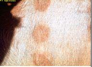 |
| Early skin lesions |
|
© F. Glynn Davies Reproduced from the Animal Health and Production Compendium, 2007 Edition. CAB International, Wallingford, UK, 2007.
|
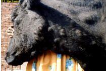 |
| Late skin lesions |
|
© F. Glynn Davies Reproduced from the Animal Health and Production Compendium, 2007 Edition. CAB International, Wallingford, UK, 2007.
|
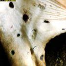 |
| Healing skin lesions |
|
© F. Glynn Davies Reproduced from the Animal Health and Production Compendium, 2007 Edition. CAB International, Wallingford, UK, 2007.
|
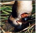 |
|
© F. Glynn Davies Reproduced from the Animal Health and Production Compendium, 2007 Edition. CAB International, Wallingford, UK, 2007.
|
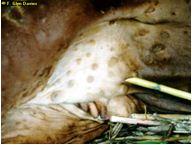 |
| Lesions on the udder |
|
© F. Glynn Davies Reproduced from the Animal Health and Production Compendium, 2007 Edition. CAB International, Wallingford, UK, 2007.
|
|
|
|
Damaged hide after tanning |
|
© F. Glynn Davies Reproduced from the Animal Health and Production Compendium, 2007 Edition. CAB International, Wallingford, UK, 2007.
|
- Animals may salivate profusely. A clear discharge comes from the eyes and nose. Later the discharge from the nose becomes grayish/white.
- The cattle are weak and tired and stop eating. They have a fever that sometimes goes down after 1 - 2 days but it goes up again. Animals produce little milk and pregnant cattle often abort.
- Lumps appear on the body, usually around the head and neck, under the abdomen, on the legs, or around the genitals and the udder. Sometimes the whole body is covered in lumps.
- The lumps are hard and usually all are about the same size- from 0.5 to 5.0 cms in diameter. The hair on the lumps stands up. The lumps are located within the skin and are firm, raised, round and flattened. Softer, yellowish/grey lumps may appear on the mouth. They rub off easily leaving sore red patches.
- The lymph nodes enlarge and sometimes a leg swells with persistent, painful oedema, accompanied by sloughing of the skin, leaving a large, open suppurating wound. Scars may be left which damage the hide. Healing may take several weeks, and hardened lumps may remain.
- The nodules dry and slowly begin to separate from the surrounding skin, finally sloughing, leaving an unsightly ulcer, which slowly heals, leaving behind a damaging scar.
- Adult cattle do not usually die but they may take months to recover and a few of them become very thin.
- Young calves often die.
- Occasionally the disease is very mild; animals only have a low fever and lumps in the skin that heal in about six weeks.
- Sometimes nodules spread to the upper respiratory tract, causing difficulty in breathing, and death within 10 days.
- Sometimes many animals are affected, sometimes only a few. Of those affected, some animals are very ill and may die. Others are only mildly ill. Yet others may take a long time to recover, and remain in poor condition for weeks. Pregnant cows may abort.
The sudden appearance of lumps in the skin after an initial fever, with sometimes rapid spread to other animals make it difficult to confuse Lumpy Skin Disease with any other disease. The only other disease which might cause confusion - Pseudo-lumpy Skin Disease (PLSD) - is only seen on the outside layers of the skin, or confined to the teats.
PLSD
The PLSD virus primarily causes Bovine Ulcerative Mammillitis, a severe, ulcerative condition of the teats and udder of cows, mostly newly calved dairy heifers, but also previously unexposed cows. Biting insects are believed to initiate infection.
Signs of PLSD:
- PLSD, like true LSD, causes circular or oval plaques in the skin that are about 1cm in diameter, are hard and firm and have a red edge.
- Over the next few days the plaques enlarge to a diameter of 3-5cm. Their centres are depressed, discharge and form a thin brown crust.
- The skin beneath the crust dies and peels off 2 weeks later, leaving a bald patch of new skin covered with fine, grey scales. New hair covers the area within 2 months
- The number of plaques ranges from a few to many and they may be found anywhere on the skin, but mostly on the face, neck, back and perineum.
This condition is endemic in Eastern and Southern Africa and the US. This is a benign disease for which treatment is not warranted and is often missed. Its importance lies in its superficial similarity to true Lumpy Skin Disease.
LSD Mode of spread
Outbreaks of true Lumpy Skin Disease often occur at the start of the rains. Although biting insects are believed to be mainly responsible for transmission, contact infection via infected saliva is also accepted as a method of spread.
Known sources of infection include permanent swamps, from where new outbreaks may start. Outbreaks may be small and isolated or gradually spread to cover large areas of the country and even spread into neighbouring countries, affecting thousands of animals. Imported breeds of cattle appear to be more susceptible than local breeds. The disease may be confused with besnoitosis, dermatophilosis, or ringworm.
The incubation period ranges from 2 to 4 weeks. The disease may be peracute (very rapid onset), acute (rapid onset) or inapparent. All ages are affected.
Prevention and control
Vaccination for Lumpy Skin Disease is effective. Vaccinate all animals in contact with the disease. Preventive vaccination is also advisable using the modified sheep/goat pox vaccine made by KEVEVAPI in Nairobi.
PLSD is of relative insignificance other than that it can confuse the diagnosis of the much more significant LSD.
Recommended treatment
There is no specific treatment for Lumpy Skin Disease but the prevention of secondary infection is vital, using antibiotics and sulphonamides.

